3D Imaging Reveals How Mosquitoes Detect Carbon Dioxide With Specialized Neurons
Scientists at the University of California San Diego (UCSD) have uncovered in remarkable detail how mosquitoes are able to detect the carbon dioxide (CO₂) that humans and animals exhale—a key part of how these insects track us down for their blood meals. Using advanced 3D electron microscopy, researchers reconstructed the mosquito’s CO₂-sensing neurons at the nanoscale, offering the most detailed look yet at the inner architecture that makes mosquitoes such efficient hunters.
This new research, published in the Proceedings of the National Academy of Sciences (PNAS), reveals intricate anatomical adaptations within the olfactory receptor neurons (ORNs) located in the sensory hairs of mosquitoes. The study provides a clear visual and structural explanation for something scientists have known for decades but couldn’t fully understand: how mosquitoes are so good at finding humans.
A Close Look at the Mosquito’s CO₂ Detection System
Mosquitoes locate their hosts using multiple cues—body heat, skin odor, and particularly carbon dioxide from exhaled breath. When you breathe out, even a small puff of CO₂ in the air can act as a signal that there’s a living host nearby.
The UCSD team focused on Aedes aegypti, the mosquito species responsible for transmitting yellow fever, dengue, chikungunya, and Zika virus. These insects are among the most dangerous in the world, partly because their sensory systems are highly evolved for finding human hosts.
At the National Center for Microscopy and Imaging Research on the UCSD campus, the scientists used a technique called serial block-face electron microscopy (SBEM). This involves slicing extremely thin layers of tissue and capturing detailed electron-beam images of each layer. By stacking thousands of these images together, the team was able to create a 3D nanoscale model of the mosquito’s CO₂-sensing neurons—something that had never been visualized before.
What the 3D Models Revealed
Inside the mosquito’s sensory hairs, known as sensilla, lie clusters of olfactory receptor neurons. Each of these hairs contains three different neuron types: cpA, cpB, and cpC. Among them, the cpA neuron is specialized for detecting CO₂.
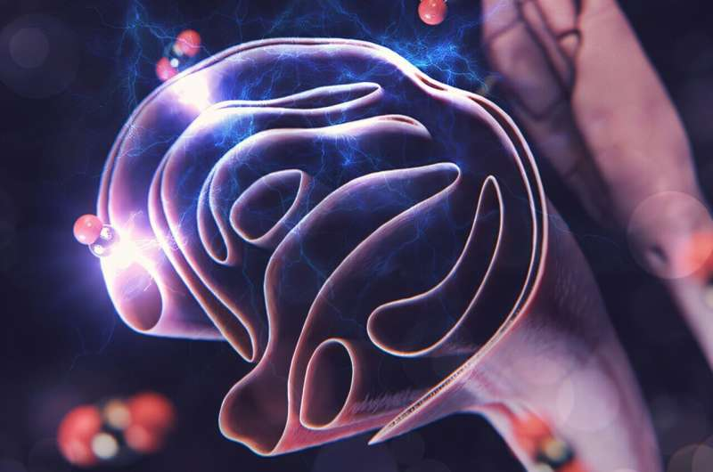
The 3D reconstructions revealed that these cpA neurons possess dramatically enlarged surface areas, up to 8 to 12 times greater than the other neurons in the same hair. This massive surface expansion comes from flattened dendritic sheets—thin, leaf-like projections that increase the cell’s ability to sense gas molecules. More surface area means more receptor sites for CO₂, giving the mosquito a much higher sensitivity to even faint concentrations in the air.
The researchers also discovered that these CO₂-sensing neurons feature a unique axonal architecture filled with mitochondria, the energy-producing structures inside cells. This suggests that the neuron’s CO₂ detection process is highly energy-intensive, requiring constant power to maintain its readiness to respond to changes in the environment.
Additionally, the cpA neuron is wrapped by a support cell, forming a kind of insulated compartment. This insulation may help stabilize the neuron’s environment and preserve signal precision. Interestingly, this surrounding support layer wasn’t found in similar neurons in other insects.
Comparison With Fruit Flies
To understand what makes mosquitoes so specialized, the team compared their results to fruit flies (Drosophila), which also have CO₂-sensing neurons. In fruit flies, however, the CO₂-sensing area is much smaller and structurally simpler.
This difference lines up perfectly with each species’ lifestyle. Fruit flies don’t feed on blood, so CO₂ acts as a warning signal—it tells them to fly away, since it often means danger (like approaching mammals). For mosquitoes, though, CO₂ means food nearby. It’s an arousal cue that activates their host-seeking behavior, prompting them to home in on whoever exhaled it.
This contrast between the two insects helps explain why mosquito sensory neurons have evolved such specialized structures. Their survival depends on their ability to detect CO₂, while for fruit flies, it’s just another odor to avoid.
Why These Findings Matter
Mosquitoes are often called the deadliest animals on Earth, responsible for hundreds of thousands of deaths every year due to the diseases they spread. Understanding how they find and target humans could help scientists design better repellents or traps that disrupt these detection mechanisms.
For instance, knowing that the cpA neuron has an unusually large surface area and a distinct energy-demanding architecture could inspire the development of chemical or molecular inhibitors that interfere with its function. Blocking or confusing the mosquito’s CO₂ detection system might keep them from recognizing us as potential hosts, reducing bites and disease transmission without relying entirely on insecticides.
Moreover, the research emphasizes that these sensory systems have evolved with high precision. Every structural feature—from the lamellar dendritic sheets to the mitochondrial enrichment—contributes to a heightened ability to detect even trace amounts of carbon dioxide.
This study gives scientists a concrete structural foundation for understanding how the mosquito’s brain receives and processes CO₂ information—something that has been largely theoretical until now.
Understanding the Broader Context of Mosquito Sensory Biology
Mosquitoes use a combination of senses to find hosts, including:
- Olfactory cues: detecting odors like lactic acid, ammonia, and CO₂.
- Thermal cues: sensing body heat from several meters away.
- Visual cues: recognizing dark colors and motion, particularly in daylight.
Their maxillary palps—tiny appendages near the mouthparts—contain the specialized sensilla where these CO₂-sensitive neurons reside. The olfactory receptor genes in these neurons code for proteins that bind CO₂ molecules, triggering electrical signals to the mosquito’s brain.
Once a mosquito senses CO₂, it becomes more active and alert, starting to use its vision and thermal sensors to zero in on its target. This makes CO₂ detection the first and most important step in the chain of behaviors that lead to a bite.
Understanding these combined systems is vital for public health because it highlights multiple points where mosquito detection can potentially be disrupted. For example, researchers have already been studying gene-editing techniques to knock out or modify specific olfactory receptors in Aedes aegypti, aiming to make them less effective at finding humans.
The Technology Behind the Discovery
The imaging technology used in this study—serial block-face scanning electron microscopy—is one of the most advanced forms of 3D microscopy available. It can produce nanoscale resolution, meaning it captures structures at the level of billionths of a meter.
Each round of imaging involves cutting off a thin layer of tissue, taking an electron micrograph, then repeating the process thousands of times. The resulting image stack is digitally reconstructed into a 3D model, allowing researchers to rotate, measure, and analyze fine details of neuronal anatomy that can’t be seen in standard microscopes.
Using this technology, the UCSD team was able to quantify the surface area of dendrites, map mitochondrial distribution, and visualize neuron–support cell relationships. This approach provided both qualitative and quantitative insights into how these neurons are built to function.
What Comes Next
While this study focused on mosquito anatomy, it opens the door to many new questions. Could manipulating the CO₂-sensing pathway stop mosquitoes from recognizing humans altogether? Can similar structural features be found in other disease-vector species, like Anopheles, which spread malaria?
Future work could involve combining these 3D imaging results with electrophysiological recordings—measuring how these neurons actually respond to CO₂ in real time. Such experiments could reveal how structure translates to sensitivity, and whether blocking energy metabolism in the cpA neuron affects its function.
As research continues, these findings might inform new mosquito-control strategies that target sensory biology rather than relying purely on pesticides, potentially offering more sustainable and species-specific solutions.
Study Details
The study, titled “Morphological Specializations of Mosquito CO₂-Sensing Olfactory Receptor Neurons”, was led by undergraduate researchers Shadi Charara and Jonathan Choy in the lab of Professor Chih-Ying Su at UC San Diego’s Department of Neurobiology. Collaborators included scientists from UCSD’s School of Medicine, the National Center for Microscopy and Imaging Research, and the Department of Cell and Developmental Biology.
The research team combined biological imaging, quantitative morphology, and comparative analysis with fruit flies to produce a comprehensive look at the mosquito’s CO₂-detection apparatus. Their work provides a visual and measurable basis for understanding one of the most critical sensory systems in vector biology.
Research Paper: Morphological Specializations of Mosquito CO₂-Sensing Olfactory Receptor Neurons – Proceedings of the National Academy of Sciences (2025)
