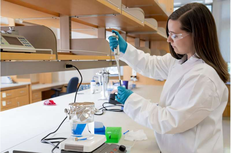
Engineers Develop Conductive Bioelectronic Hydrogels That Can Monitor Activity Inside the Body

Scientists at Washington University in St. Louis have taken a big step toward creating soft, injectable, and flexible electronics that could one day work seamlessly with our bodies. Instead of relying on metals, silicon, or rigid plastics, their new technology uses bioelectronic hydrogels—soft, conductive materials that can monitor and even stimulate biological activity inside living tissues.
The research, led by Assistant Professor Alexandra Rutz and her doctoral student Anna Goestenkors, was recently published in the journal Small on October 8, 2025. Their work introduces a new kind of granular hydrogel made from tiny conductive particles that can be injected into the body, spread over tissues, or even 3D printed into customized shapes.
What Are Bioelectronic Hydrogels?
Hydrogels are materials made mostly of water and polymers that hold it in a soft, gel-like structure. Because they are wet and flexible, they resemble biological tissues. Traditionally, hydrogels have been used for wound dressings, contact lenses, tissue scaffolds, and even drug delivery systems.
What makes this research special is that the hydrogels developed by the WashU team are also electrically conductive. That means they can form an interface between electronics and living tissues, something that is incredibly difficult to achieve.
The team achieved this by using a conducting polymer called PEDOT:PSS, a well-known material in bioelectronics. PEDOT:PSS can transport electrical signals while remaining flexible and biocompatible—qualities that are ideal for devices that must work inside or closely with the body.
How the Material Works
The hydrogels aren’t just one solid block. Instead, they are made from tiny spherical microparticles—each one a miniature hydrogel bead about 10 to 100 micrometers in diameter. When packed together, these particles form what is known as a granular hydrogel.
The key advantage of this structure is adaptability. When these microparticles are packed tightly, they behave like a soft solid—a paste that holds its shape. But when force is applied, such as during injection through a needle or extrusion through a 3D printer, the particles can slide past one another, behaving like a liquid. Once the force is removed, they settle back into a semi-solid state.
This reversible behavior is known as shear-thinning. It makes the material ideal for injection into tissue or for 3D printing soft electronic devices that need to match the contours of organs or skin.
The microparticles themselves are created using a water-and-oil emulsion process. The polymer solution (PEDOT:PSS) is mixed with oil and stirred, similar to how one might whisk oil and vinegar into a salad dressing. When the mixture is heated, the polymer forms crosslinked, stable hydrogel particles suspended in the oil. Once isolated and cleaned, these particles can be assembled into a conductive gel.
Because of the micropores between the particles, the hydrogel also allows cells, nutrients, and fluids to move through it—something rigid materials can’t do. This means it can serve as a medium not only for electrical monitoring but also for cell growth and tissue integration.
Why This Is a Breakthrough
Most wearable or implantable medical devices—like pacemakers or neural electrodes—are built from hard materials such as metals and silicon. They work well electronically, but they don’t “match” the body. Tissues are soft, elastic, and constantly moving. Rigid devices can cause inflammation, scarring, or mechanical failure over time.
The granular hydrogel developed by the Rutz lab could solve that problem. It is soft, stretchable, and conductive, behaving more like living tissue while still allowing electronic communication.
Unlike traditional electrodes that need surgical placement, this hydrogel could potentially be injected with a simple needle, conforming to the shape of the target area without invasive surgery. It could also be 3D printed directly onto tissues during surgery or lab-grown tissue fabrication.
How It Was Tested
To prove that the material can actually record biological signals, the researchers conducted an experiment with locusts. In collaboration with Professor Barani Raman, who co-directs WashU’s Center for Cyborg and BioRobotic Research, they placed small clumps of the hydrogel microparticles on the tips of locust antennae.
These antennae contain olfactory receptor neurons, which generate electrical signals when they detect odors. The hydrogel particles successfully captured local field potentials corresponding to the locusts’ sensory responses. In simple terms, the material was able to “listen” to the brain’s electrical signals from a living organism.
This experiment demonstrates that the granular hydrogel can not only exist comfortably on biological tissue but also communicate with it electrically. That’s a big leap forward in developing flexible, injectable bioelectronic interfaces.
Potential Applications
The possibilities for this technology are exciting and wide-ranging. Some potential applications include:
- Soft bioelectronic implants that monitor neural or cardiac activity without damaging tissue.
- Injectable sensors for real-time monitoring of internal processes such as muscle activity or nerve signals.
- 3D printed electrodes that conform to complex organ shapes or wrap around soft tissues.
- Tissue engineering scaffolds that can both support growing cells and measure their activity.
- Targeted electrical stimulation of nerves or muscles for therapeutic purposes.
In essence, this material could lead to a new generation of soft, adaptive medical devices that feel more like part of the body than something implanted into it.
Challenges and Next Steps
Even though the results are promising, the technology is still in the early stages. Several challenges remain before it can be used in humans:
- Long-term biocompatibility needs to be studied to ensure the material doesn’t trigger immune reactions or degrade unpredictably inside the body.
- Mechanical stability over weeks or months of movement must be verified, especially in organs like the heart or brain.
- The team will also need to test signal reliability and resolution, ensuring that the soft hydrogel can transmit clear, accurate readings comparable to traditional electrodes.
- Manufacturing and scalability could also pose difficulties. Creating uniform microparticles and assembling them reproducibly at scale will require engineering optimization.
The research team is already working with WashU’s Office of Technology Management to patent the fabrication method and its applications. This is an important step toward commercial development.
Understanding PEDOT:PSS — The Conductive Polymer Behind the Breakthrough
At the heart of this innovation is PEDOT:PSS, short for poly(3,4-ethylenedioxythiophene):poly(styrenesulfonate). It’s a mouthful, but here’s why it’s so important.
PEDOT:PSS is one of the most widely used conductive polymers in organic electronics. It combines electrical conductivity with mechanical flexibility—qualities that traditional metal-based electronics lack.
You’ll find PEDOT:PSS in applications like:
- Flexible touchscreens and displays
- Organic solar cells
- Smart textiles and bioelectronic sensors
In bioelectronics, it’s particularly valuable because it’s biocompatible and can operate in wet environments—like the human body. The WashU team’s approach takes PEDOT:PSS to a new level by transforming it into a granular hydrogel format, giving it a unique combination of softness, conductivity, and injectability.
How Granular Hydrogels Differ from Traditional Hydrogels
Traditional hydrogels are continuous networks of polymer chains swollen with water. They’re soft and flexible but not easily reconfigurable once formed. Granular hydrogels, on the other hand, are composed of discrete microparticles that interact physically rather than chemically.
This design gives them several advantages:
- Self-healing: The particles can move and reconnect after being disturbed.
- Injectability: They can flow under pressure and re-solidify after injection.
- Porosity: Gaps between the particles allow nutrient and cell transport.
- Customizability: They can be 3D printed into complex shapes or blended with other materials.
In essence, granular hydrogels combine the mechanical tunability of granular matter with the biocompatibility of hydrogels—a hybrid that’s perfect for bioelectronic applications.
The Broader Context: Soft Bioelectronics and the Future of Medical Devices
This research fits into a rapidly growing field known as soft bioelectronics. The goal is to create electronic devices that move, stretch, and feel like biological tissue, reducing mechanical mismatch and improving long-term performance inside the body.
Recent advances include:
- Stretchable electronic tattoos that can record heart or muscle activity directly from the skin.
- Flexible neural interfaces that can record brain activity without damaging neurons.
- Soft electronic scaffolds that help regrow damaged nerves or muscles while monitoring progress.
The WashU team’s granular hydrogel adds something new to this landscape: injectability and 3D printability, which could dramatically simplify how electronic interfaces are deployed in living tissue.
Imagine a future where doctors can inject a soft, conductive gel directly into a damaged nerve or heart tissue, and it instantly forms a functioning sensor or stimulator. That’s the kind of potential this technology is hinting at.
Final Thoughts
The creation of these conductive granular hydrogels marks a fascinating convergence of biology, materials science, and electronics. By using PEDOT:PSS microparticles to form a soft, injectable, and reconfigurable material, the team at Washington University has opened a new pathway for building medical devices that can truly integrate with the human body.
While more research is needed to bring this technology from the lab to clinical use, its implications are hard to ignore. From wearable health monitors to implantable neural interfaces, this kind of soft, biofriendly material could redefine how humans and machines connect.
Research Reference:
PEDOT:PSS Microparticles for Extrudable and Bioencapsulating Conducting Granular Hydrogel Bioelectronics – Small (2025)


